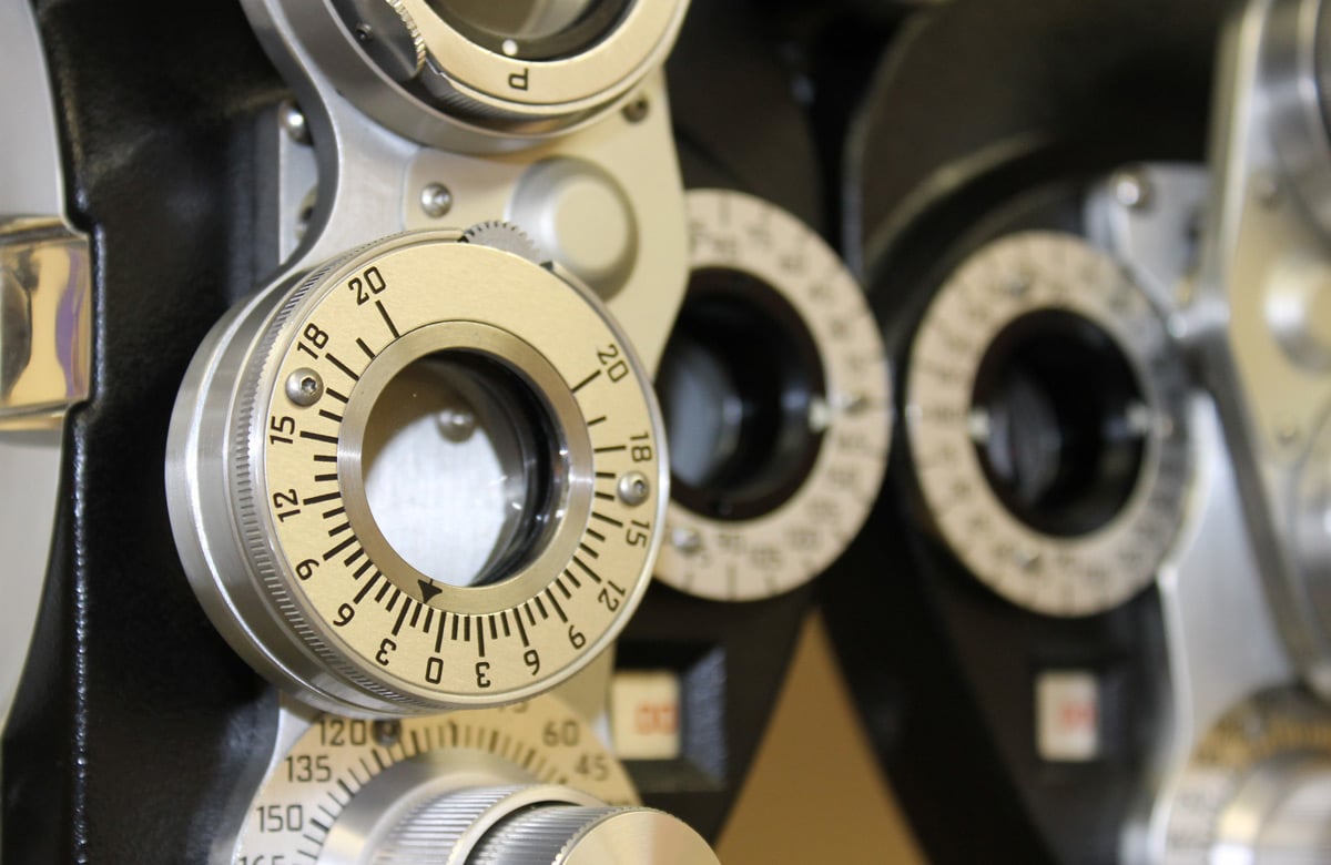
Developed by bioengineering researchers from the University of Maryland (UMD), the microscopy technique would allow doctors to perform LASIK procedures based on precise measurents of how the eye focuses light, rather than approximations and guesswork“This could represent a trendous first for LASIK and other refractive procedures,” assistant professor Giuliano Scarcelli, study lead at the UMD Fischell Department of Bioengineering (BIOE), said.“Light is focused by the eye’s cornea because of its shape and what is known as its refractive index. But until now, we could only measure its shape. Thus, today’s refractive procedures rely solely on observed changes to the cornea, and they are not always accurate.”Together with his research team at the Optics Biotech Laboratory, Scarcelli has developed a technique that can measure the local refractive index using Brillouin spectroscopy—a light-scattering technology that was previously used to detect the mechanical properties of tissue and cells without disrupting or destroying th.“This means that we could measure the refractive index of cells and tissue at locations in the body—such as the eyes—that can only be accessed from one side, Scarcelli said.Equipped with the precise degree of corneal refraction, the team hopes that doctors could tailor a LASIK procedure to allow a patient to walk away with perfect vision, rather than just improved vision. It may even make cutting into the cornea unnecessary.The study was published in the journal Physical Review Letters.
BHVI help Haiti launch country?s first optometry school
A collaboration involving the Brien Holden Vision Institute opened Haiti’s first optometry school last month, in a positive step for...


